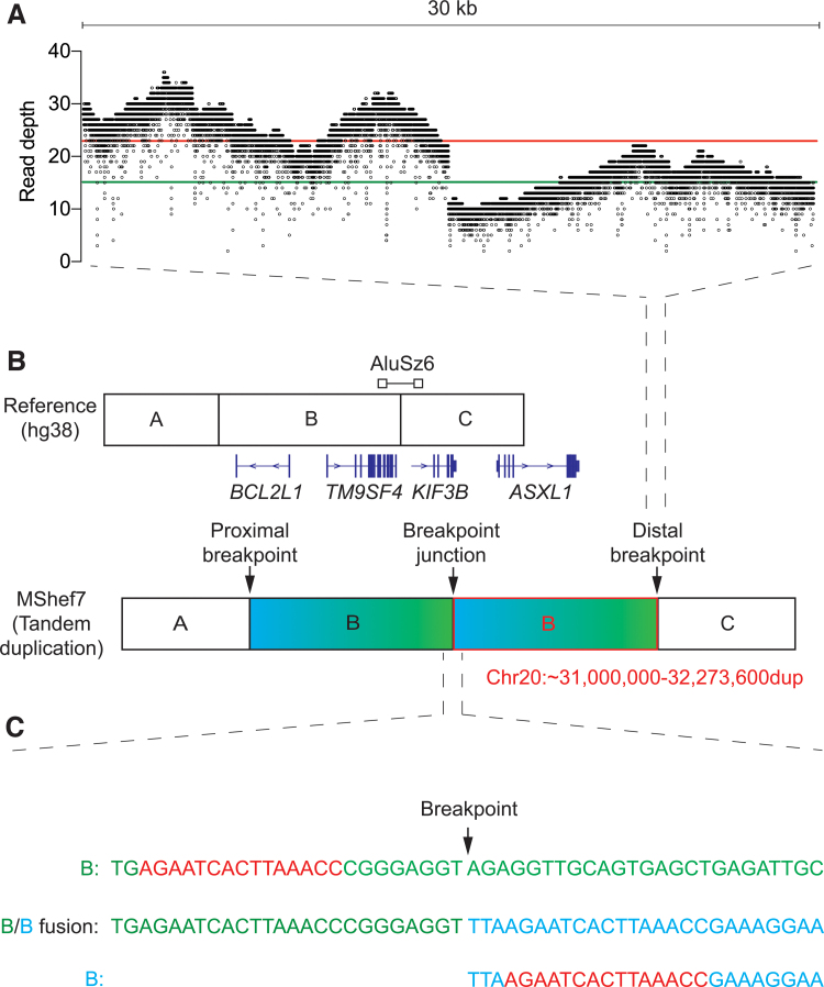
FIG. 2.
Breakpoint junction detection in MShef7-A4 using Nanopore sequencing. (A) Sequencing read coverage of 30 kb spanning the distal breakpoint junction at 32,273,600 bp (chromosome 20q11.21) of the hg38 reference genome. Each dot indicates the read depth at a single base pair position. The red and green lines indicate the mean read depth before and after the breakpoint position, respectively. (B) Schematic of the reference genome and the tandem duplication detected in MShef7-A4. Junction between genome segment A-B and B-C represents the proximal and distal breakpoints, respectively. The position of genes flanking and the location of the AluSz6 in relation to the breakpoint are depicted. (C) Reference sequence spanning the distal breakpoint (B—top, green), sequence of the breakpoint junction (B/B fusion—middle), and the contig sequence of the distal side of the proximal breakpoint (B—bottom, blue). The regions of microhomology that flank the proximal and distal breakpoints are indicated in red. Color images are available online.

