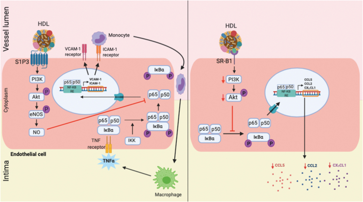
Figure 2.
Mechanisms for the anti-inflammatory effects of HDL in endothelial cells. Left panel: HDL increase formation of bioactive nitric oxide (NO) through interaction with the S1P3 receptor, which activates PI3K/Akt, leading to inhibition in action of NF-κB. This prevents the translocation of the NF-κB subunits p65/p50 to the nucleus, where it binds to the NF-κB response elements (RE) in the promoter region of inflammatory genes, including vascular cell adhesion molecule (VCAM)-1 and intercellular cell adhesion molecule (ICAM)-1. Right panel: in an alternative pathway, HDL reduce the production of inflammatory cytokines CCL2, CCL5, and CX3CL1 through interaction with scavenger receptor (SR)-B1 that leads to suppression of downstream kinase signaling (PI3K/Akt) and inhibition in the activation of NF-κB pathway. NF-κB, nuclear factor kappa B; S1P3, sphingosine-1-phosphate 3. Color images are available online.

