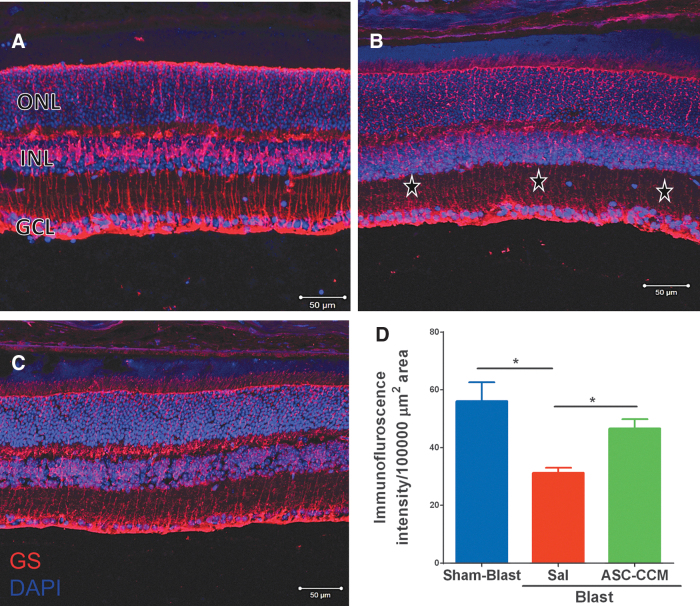
FIG. 2.
Decrease in glutamine synthetase (GS) was seen in blast injury retina and improved by intravitreal injection of adipose tissue–derived mesenchymal stem cell concentrated conditioned medium (ASC-CCM). Immunohistological analysis of retinal tissue from all groups 4 weeks post–blast injury. Confocal micrographs showing GS (red) in the retina of Sham-Blast (A), Blast-Sal (B), and Blast/ASC-CCM (C) with 4′,6-diamidino-2-phenylindole (DAPI; blue) staining showing different retinal layers (GCL, ganglion cell layer; INL, inner nuclear layer; ONL, outer nuclear layer). (D) Image J quantification of GS intensity. * represent areas of significant change. Data represent mean ± SEM from n = 6 animals/group (Sham-Blast vs. Blast-Sal **p < 0.003, Blast-Sal vs. Blast/ASC-CCM *p < 0.049). Scale bar = 50 μm. Color image is available online.

