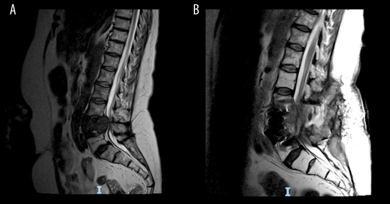
Figure 1.
(A) Sagital T2-weighted magnetic resonance imaging (MRI) of lumbar-sacral spine showing cauda equina compression at L4 level at first presentation of differentiated thyroid cancer. (B) Sagital T2-weighted MRI of lumbar spine showing progressive disease, complex abscess formation, and cauda equina compression at L4 level at point of anaplastic thyroid cancer transformation.

