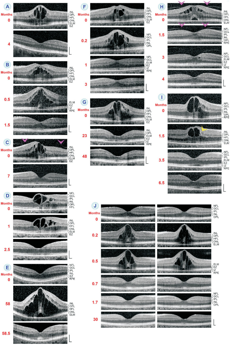
Figure 1. Regeneration of the foveal morphology after resolution of cystoid macular edema (CME).
The images show SD-OCT scans through the fovea and parafovea of 11 eyes of 10 patients. The months after the first visit (0) are indicated left of the images. A-G: Morphological regeneration of the fovea after resolution of CME in eyes of different patients. Arrowheads indicate adhesions of the partially detached posterior hyaloid at the para- and perifovea, respectively. H, I: Two eyes with CME with different patterns of edematous cyst distribution, characterized by the absence (H) and presence (I) of a large cyst in the foveola. H: The CME was likely induced by traction of the partially detached posterior hyaloid which adhered at the parafovea (arrowheads). I: Note that the stalk of the Müller cell cone is stretched and elongated. The arrowhead points to hyperreflective dots which represent light reflections at exudates in the inner nuclear layer (INL). J: Simultaneous CME in the foveas of the left (left side) and right eyes (right side) of a patient with uveitis which was treated with systemic prednisolone pulse therapy. The CME resolved rapidly during the subsequent systemic immunosuppressive therapy. Scale bars, 200 µm. ELM: External limiting membrane; EZ: Ellipsoid zone; GCL: Ganglion cell layer; HFL: Henle fiber layer; IPL: Inner plexiform layer; IZ: Interdigitation zone; NFL: Nerve fiber layer; ONL: Outer nuclear layer; OPL: Outer plexiform layer; RPE: Retinal pigment epithelium.

