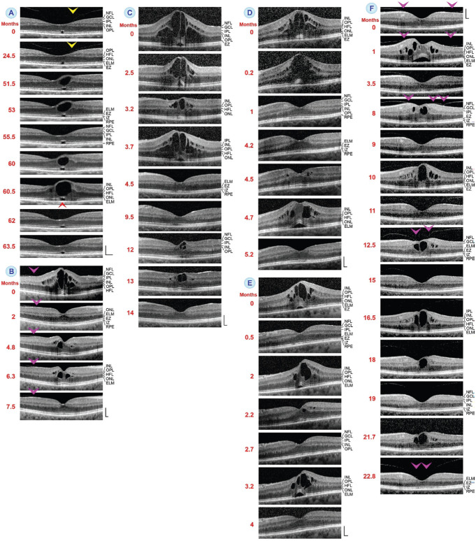
Figure 2. Regeneration of the foveal morphology after twofold (A-D) and multiple episodes (E, F) of cystoid macular edema (CME).
The images show SD-OCT scans through the fovea and parafovea of 6 eyes of 6 patients. The months after the first visit (0) are indicated left of the images. A: Twofold development and resolution of an edematous cyst in the foveola. The yellow arrowheads point to adhesion of the partially detached posterior hyaloid. The red arrowhead indicates the glial tissue membrane at the external limiting membrane (ELM) which keeps the central outer fovea together. B: The CME was likely associated with traction of the partially detached posterior hyaloid which adhered at the parafovea (arrowheads). C: Note the irregular reflection intensities of the central ellipsoid zone (EZ) and interdigitation zone (IZ) lines during both episodes of CME which were abrogated at the end of the examination period. D: An eye with different patterns of edematous cyst distribution in both episodes of CME. E: An eye with three CME episodes associated with a detachment of the central outer fovea from the RPE and subsequent regeneration of the foveal morphology. F: An eye with sixfold development of CME. The arrowheads point to foveal adhesions of the partially detached posterior hyaloid. Scale bars, 200 µm. GCL: Ganglion cell layer; HFL: Henle fiber layer; INL: Inner nuclear layer; IPL: Inner plexiform layer; NFL: Nerve fiber layer; ONL: Outer nuclear layer; OPL: Outer plexiform layer; RPE: Retinal pigment epithelium.

