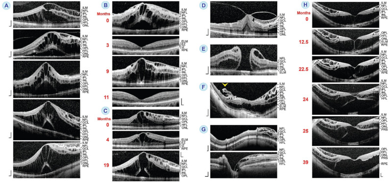
Figure 3. Detachment of the internal limiting membrane (ILM) from the nerve fiber layer (NFL) in different retinal disorders.
The images show SD-OCT scans through the fovea and parafovea of 12 eyes of 12 patients. The months after the first visit (0) are indicated left of the images. A: Five eyes with cystoid macular edema (CME) and ILM detachment in the tissues at the right side. The images also display a foveoschisis between the outer plexiform layer (OPL) and Henle fiber layer (HFL), cystic cavities in the ganglion cell layer (GCL) and inner nuclear layer (INL), and in four eyes a detachment of the fovea from the retinal pigment epithelium (RPE). In three eyes, the GCL is degenerated. The tissues at the left side of three eyes show no abnormalities with the exception of a large foveoschisis in the HFL. B: An eye with two episodes of CME and ILM detachment. C: Enlargement of an outer lamellar hole (OLH) with ILM detachment and foveoschisis between the OPL and HFL in an eye with CME. D: Fovea of an eye with macular pucker with ILM detachment in the para- and perifovea at the right side. E: An eye with a full-thickness macular hole with ILM detachment in the foveal walls and parafovea. F: An eye with high myopia. The fovea (middle) and wide retinal areas are split by a schisis within the HFL. In addition, there is ILM detachment and schistic cavities in the GCL and INL of the inferior perifovea (right side). In the superior perifovea (left side), the edge of the thickened ILM and parts of the NFL are detached from the remaining NFL (arrowhead). G: An eye with glaucomatous parapapillary retinoschisis in the nasal retina. The image above shows a circumpapillary SD-OCT scan. The image below shows a horizontal SD-OCT scan through the excavated optic disc. H: Development of an OLH with inner and outer retinoschisis in an eye with myopic traction maculopathy. Scale bars, 200 µm. ELM: External limiting membrane; EZ: Ellipsoid zone; IPL: Inner plexiform layer; IZ: Interdigitation zone; ONL: Outer nuclear layer; PRS: Photoreceptor segments; RPE: Retinal pigment epithelium.

