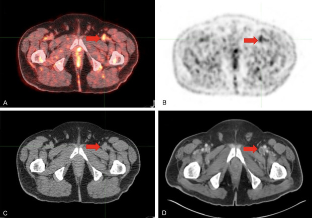
Figure 1.
Venous thrombosis in the left common femoral vein of a 65-year old male with a history of metastatic melanoma. One day prior to FDG-PET/CT imaging, the patient had +1 edema of the left calf and foot. (A) Axial FDG-PET/CT, (B) axial FDG-PET, and (C) axial low-dose CT confirm high metabolic activity in the dilated lumen of the left common femoral vein, consistent with venous thrombosis. (D) Six-month follow-up, abdominopelvic contrast-enhanced CT scan showed a filling defect in the same location, suggestive of venous thrombosis.

