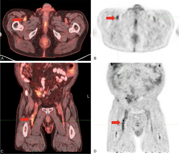
Figure 2.
Venous thrombosis in the right common femoral vein, diagnosed prior to the ultrasound. (A) Axial FDG-PET/CT, (B) axial FDG-PET, (C) coronal FDG-PET/CT, and (D) coronal FDG-PET demonstrated high metabolic activity in the lumen of the right common femoral vein, consistent with venous thrombosis. Ultrasound performed two weeks later confirmed non-occlusive thrombus in the right common femoral vein.

