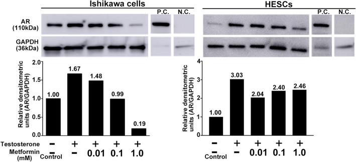
Fig. 4.
Effect of metformin on AR expression in Ishikawa cells and HESCs examined by western blotting. Representative images show AR expression by western blotting in Ishikawa cells and HESCs in the upper panel. Mouse testicular lysate was used as a positive control, and no primary AR antibody was added in the negative control. Graphs in the lower panel show the relative densitometric units (AR/GAPDH). The value in each column is the relative ratio when the control (without both testosterone and metformin) was set at 1.0. Three independent experiments showed the same results. AR, androgen receptor; HESCs, human endometrial stromal cells; N.C., negative control; P.C., positive control

