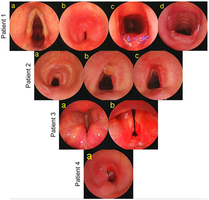
Figure 1.
Laryngotracheal stenosis. Patient # 1: (a) endoscopic view of the larynx and trachea, (b) severe grade tracheal stenosis, (c) 10 days after single stage tracheal resection and anastomosis (TRA), and (d) 2-months after TRA. Patient # 2: (a, b) endoscopic view of A-shape deformity and granuloma, and (c) view after endoscopic treatment. Patient # 3: (a) endoscopic view of bilateral immobile vocal cords in median position and (b) severe posterior glottic stenosis. Patient # 4: (a) severe grade tracheal stenosis.

