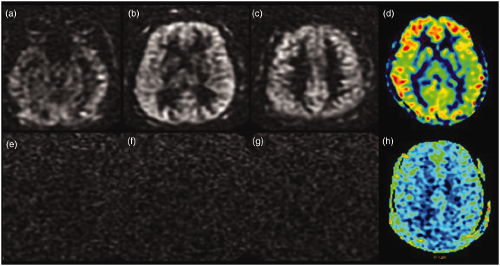
Figure 4.
Loss of spin labelling post-gadolinium injection. Serial axial tagged raw arterial spin labelling (ASL) images before the administration of gadolinium (a)–(c) and the processed colour image (d) showing good parenchymal perfusion. Source images (e)–(g) and the processed colour image (h) obtained after the administration of gadolinium-based contrast show the complete absence of ASL signal.

