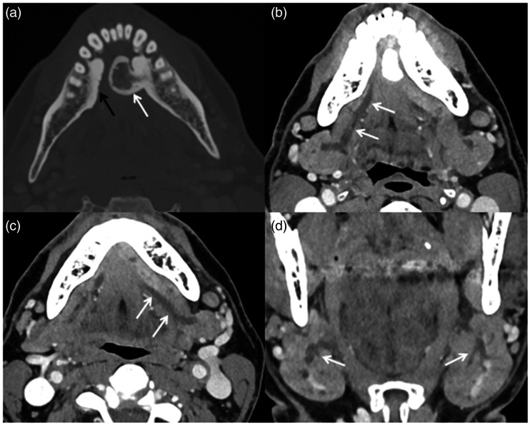
Figure 1.
Axial contrast-enhanced CT at the level of the mandible windowed for bone: (a) demonstrates a large left torus mandibularis (white arrow) and a small right torus mandibularis (black arrow). Additional images windowed for soft tissue show dilation of (b) the right (arrows) and (c) left (arrows) submandibular ducts. (d) This ductal dilation extended into both submandibular glands (arrows).

