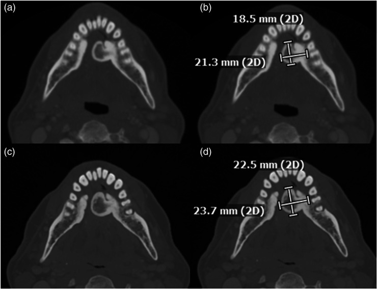
Figure 2.
Axial CT at the level of the mandible, obtained 6 years prior to the patient’s presentation: (a) without and (b) with annotated measurements, again show a left torus mandibularis. Images at the level of the mandible at the same level at the time of the patient’s presentation: (c) without and (d) with annotated measurements are shown for comparison. There is mild growth of the right torus mandibularis over the imaging interval.

