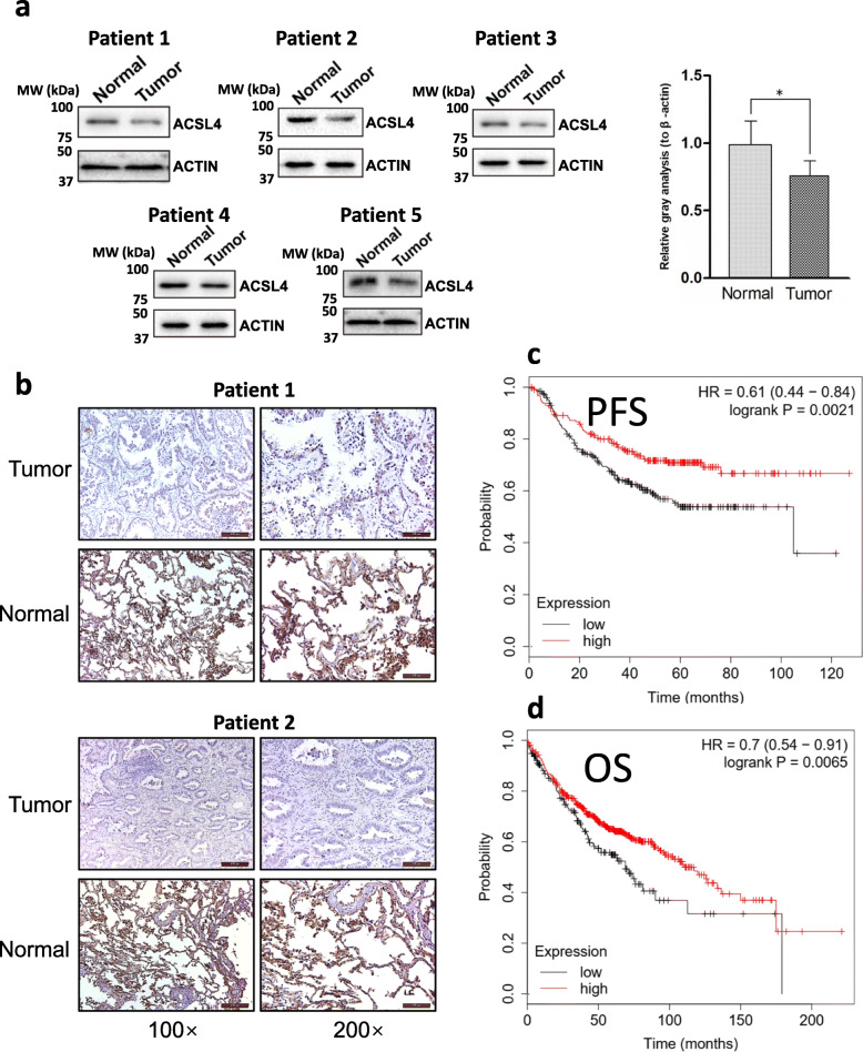
Fig. 2.
Low ACSL4 expression was associated with poor prognosis in lung adenocarcinoma. a The expression of ACSL4 in lung adenocarcinoma tissues and matched normal tissues was detected using Western blot. The samples were obtained from our hospital. Representative Western blot results were shown and ACTIN was used as the loading control. The quantification of the results (the intensity of ACSL4 divided by the intensity of ACTIN) was shown on the right. b The expression of ACSL4 in lung adenocarcinoma tissues was detected using immunohistochemistry. The samples were obtained from our hospital. c-d Kaplan-Meier analysis of progression-free survival (c) and overall survival (d) in lung adenocarcinoma patients. The patient survival data were obtained from the TCGA database (n = 524). The optimal ACSL4 expression level that was auto-selected by Kaplan Meier Plotter (see Method section for more details) was used as the cut-off to separate the high and low expression of ACSL4 patients. PFS, progression-free survival; OS, overall survival

