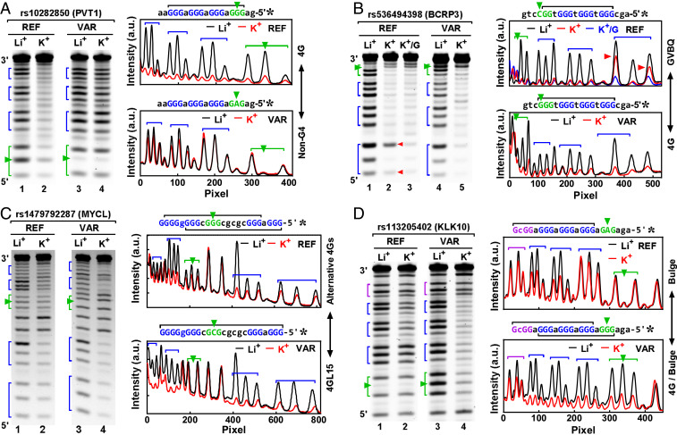
Fig. 7.
Examples of G4V caused by SNV: (A) rs10282850, (B) rs536494398, (C) rs1479792287, and (D) rs113205402. G4 formation in single-stranded DNAs was detected by the protection of the G-tracts in DMS footprinting (gels at the left side and digitization at the right side). SNV IDs are at the top of the gels, followed by the names of their host genes in parentheses. The G4 was stabilized by K+, but not by Li+. G-tracts are indicated by brackets and SNVs by arrowheads. Structural change was indicated at the right side of the digitization panel.

