Fig. 3.
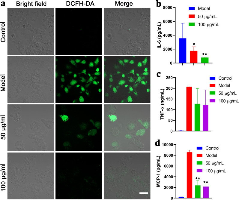
In vitro anti-oxidation and anti-inflammation using PMPB NC via scavenging ROS. a Confocal fluorescence images of RAW264.7 cells after different corresponding treatments in four groups, i.e., control, Model, 50 μg/mL, 100 μg/mL), where ROS indicator, i.e., DCFH-DA, was used to stain the intracellular ROS, scale bar: 10 μm. Herein, Model means co-stimulation of RAW264.7 cells with LPS/IFN-γ for 24 h; and the groups (50 μg/mL and 100 μg/mL) mean co-incubation of RAW264.7 cells with PMPB NC with varied concentrations (50 μg/mL and 100 μg/mL) for 2 h, followed by co-stimulation with LPS/IFN-γ for 24 h. b–d The levels of some typical inflammatory cytokines secreted by RAW264.7 cells, e.g., IL-6 (b), TNF-α (c) and MCP-1 (d), and they were detected by enzyme-linked immunosorbent assay (ELISA). Data are expressed as mean ± SD (n = 3), and *P˂0.05 and **P˂0.01, which were obtained in comparison to Model

