Fig. 1.
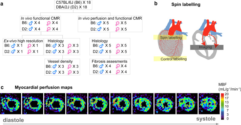
a Schematic representation of experimental design. b Schematic illustration of the tagging slice position used for quantifying myocardial blood flow (MBF) with arterial spin labelling (ASL)-cardiovascular magnetic resonance (CMR). By labelling water molecules in the inflowing blood from coronary artery as tracers which then perfused into myocardium, labelled blood signal can be imaged in the mid-left ventricle (LV). c Ten consecutive color coded short axis myocardial perfusion maps from a measurement series over one cardiac cycle

