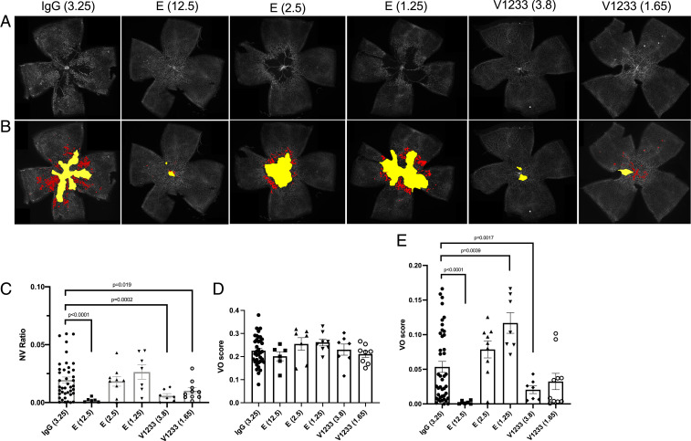
Fig. 6.
Intravitreal injections of V1233 inhibit neovascularization in the OIR model. (A) Intravitreal injections were performed at P7 in C57BL/6J mice using aflibercept (Eylea), V1233, and control (IgG). A volume of 0.5 μL aflibercept (Eylea) (E) at a dose of 12.5, 2.5, or 1.25 μg versus control IgG at a dose of 3.25 μg injected into the fellow eyes. Littermates were injected with V1233 (3.8 μg or 1.65 μg) and control. The concentrations of IgG control, aflibercept 2.5 μg, and V1233 3.8 μg were equimolar. Animals were then exposed to 75% oxygen from P7 to P12 followed by return to room air. At P17, the animals were perfusion fixed, and the eyes were enucleated, dissected, stained with lectin from Bandeiraea simplicifolia (BSL)-fluorescein isothiocyanate (FITC), and flat mounted. (B) Vasoobliteration and neovascularization were analyzed using automated software as described by Xiao et al. (72). The vasoobliterative areas are shown in yellow, and neovascular tufts are shown in red. (C) Quantification of neovascularization shows a significant reduction (P < 0.05 t test with Welch’s correction) in neovascularization relative to control with V1233 (3.8 and 1.65 μg) or high-dose aflibercept (12.5 μg), but not with aflibercept at 2.5 or 1.25 μg. (D) Quantification of vasoobliteration at P12 and (E) P17. No significant difference in VO was seen at P12 in any condition. At P17, however, a significant reduction in VO was seen with V1233 3.8 μg and high-dose aflibercept (12.5 μg), but neither with V1233 1.65 μg nor aflibercept 2.5 or 1.25 μg.

