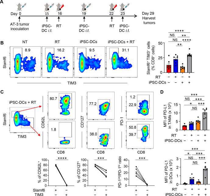
Figure 5.
A combination of in situ iPSC-DC injection and RT increases stem-like progenitor exhausted CD8+ T cells and PD-L1 expression in myeloid cells in the tumor. (A) Experimental set-up. (B) Phenotypic analysis of CD8+ T cells among CD45+ cells in AT-3 tumors in different treatment groups as indicated. Numbers denote per cent Slamf6+ TIM3− cells. Representative flow cytometric plots showing expression of Slamf6 and TIM3 in CD8+ TILs. The data panel shows the frequency of Slamf6+ TIM3− cells in CD8+ TILs (n=7). (C) Phenotypic characterization of Slamf6+ TIM3− and Slamf6− TIM3+ CD8+ TILs (n=6). (D) PD-L1 expression (MFI: median fluorescence intensity) of Ly6c− CD11c+ class II+ F4/80hi CD24− tumor-associated macrophages (TAMs) (upper) and Ly6c− CD11c+ class II+ F4/80lo CD24+ DCs (lower) in AT-3 tumors (n=7). Gating strategy for identifying TAMs and DCs is shown in online supplemental figure 3. NS not significant, *p<0.05, **p<0.01, ***p<0.001, ****p<0.0001 by one-way ANOVA with Tukey’s multiple comparisons (B, D) and two-tailed paired t-test (C). Each dot represents biologically independent mice (B, D). Data shown are representative of two independent experiments. Mean±SEM. ANOVA, analysis of variance; iPSC-DC, induced pluripotent stem cell-derived dendritic cells; i.t., intratumorally; PD-1, programmed cell death protein 1; PD-L1, PD-1 ligand 1; RT, radiotherapy; TILs, tumor-infiltrating lymphocytes; TIM3, T cell immunoglobulin and mucin domain-containing protein 3.

