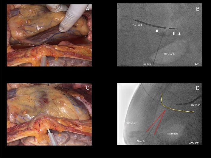
Fig. 2.
a, b Inferior epicardial access with fluoroscopy projection in AP. White arrows in b illustrate the contrast media within the pericardial space. c, d Anterior epicardial access with fluoroscopy projection in LAO 90°. Yellow dotted line illustrates the silhouette of the heart. Red dotted line illustrates the triangle between sternum, abdominal organs and heart. With permission of the Institute of Pathology, Asklepios Hospital St. George. LAO Left anterior oblique, AP Anteroposterior, RV right ventricle

