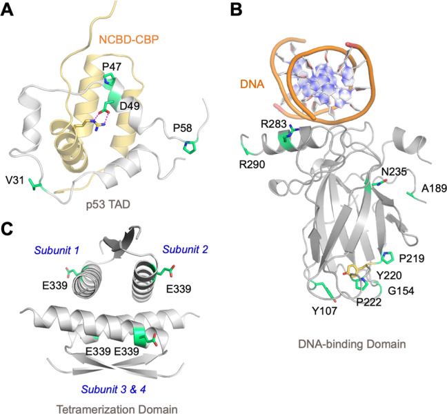
Fig. 5. Location of TP53 SNPs in the protein structure.

A Solution structure of p53 TAD in complex with the nuclear receptor coactivator binding domain of CREB binding protein (NCBD-CBP) (PDB entry 2L14). B Crystal structure of the DBD bound to target DNA (PDB entry 3KZ8, chain A). C Crystal structure of the tetramerization domain (PDB entry 1C26). p53 domain structures in A–C are shown as cartoons in gray, with the SNP sites highlighted as green stick models. NCBD-CBP is shown as a light-brown cartoon, and the side chain of the arginine involved in an intermolecular salt bridge with p53 is shown as a stick model.
