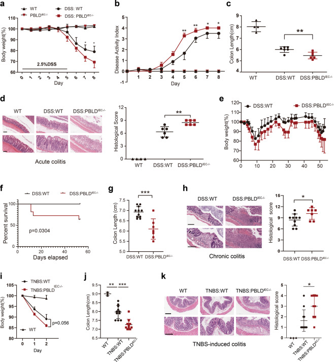
Fig. 2. Deletion of PBLD in IECs aggravates DSS- and TNBS-induced colitis in mice.
a Body weight curve and b disease activity index of wild-type (WT) and PBLDIEC−/− mice treated with DSS for 5 days, followed by 3 days regular drinking water (WT, n = 4; PBLD IEC−/−, n = 4; DSS:WT, n = 6; DSS:PBLDIEC−/−, n = 6). c Colon length of mice sacrificed at day 8. d Representative HE-stained colon sections in mice (left) and their corresponding histological score (right) (n = 4–6). Scale bars: 200 μm in upper,100 μm in lower. e Body weight curve and f survival curve in the DSS-induced chronic colitis model in WT and PBLDIEC−/− mice (n = 11 in each group). g Colon length of mice sacrificed at the end of chronic colitis model (n = 7–11). h Representative HE-stained colon sections in mice with chronic colitis (left) and their corresponding histological scores (right; n = 7–10). Scale bars: 300 μm in upper, 200 μm in lower. Data are mean ± standard deviation (SD). ***p < 0.001. i Body weight changes in WT mice and PBLD IEC−/− mice after TNBS administration. j Colon length in mice after 2 days of TNBS treatment (n = 3–11). k HE-stained colon sections in mice after 2 days of TNBS treatment (left) and their corresponding histological scores (right). Scale bars: 300 μm in upper, 200 μm in lower.

