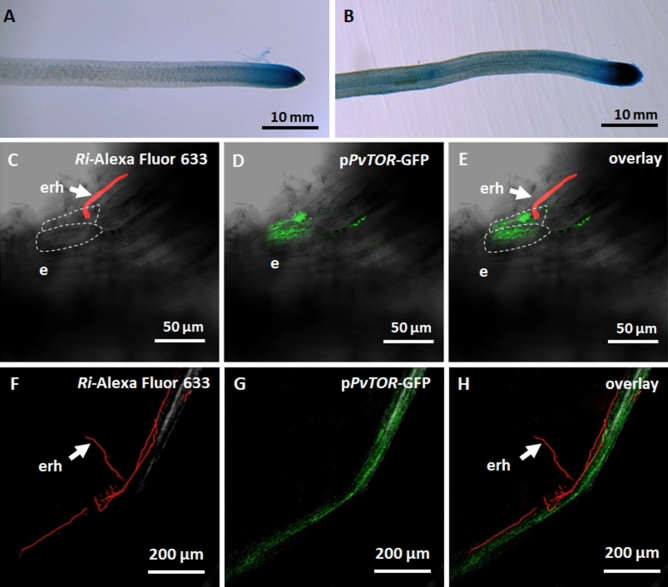
Figure 2.
Spatial expression of the PvTOR promoter in R. irregularis-inoculated transgenic P. vulgaris roots. (A–B) Histochemical GUS staining of (A) uninoculated and (B) R. irregularis-inoculated PvTORpro::GUS-GFP transgenic roots at 2 wpi. (C–E) Transmitted light and confocal images of PvTORpro::GUS-GFP transgenic roots at 2 dpi with R. irregularis showing (C) extraradical hyphae in contact with root epidermis, (D) induction of PvTORpro::GUS-GFP expression in the epidermal cells at the site of extraradical hyphal contact and (E) overlay image. (F–H) PvTORpro::GUS-GFP transgenic roots at 2 wpi with R. irregularis showing (F) extraradical hyphae on the root epidermis surface, (G) induction of PvTORpro::GUS-GFP expression in the root colonized by R. irregularis, and (H) overlay image. WGA Alexa Fluor 633 was used to stain (red) fungal cell walls. dpi, days post inoculation; wpi, week(s) post inoculation; e, epidermis; erh, extraradical hyphae.

