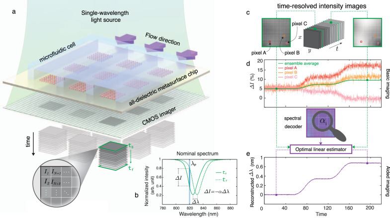
Fig. 1. Principles of the single-wavelength algorithm-aided imaging biosensor with spectral shift reconstruction and its advantages over the basic imaging approach that merely relies on the intensity change.
a Sketch of a real-time in-flow imaging platform showing a 2D microarray of all-dielectric sensors integrated with a microfluidic cell consisting of three independent flow channels. The metasurface chip is illuminated with a single-wavelength light source and imaged with a large-area CMOS camera to acquire intensity maps of the sensors in pixel resolution (I1, I2, …, IN+1, IN+2,…). b Biomarker binding induces a red-shift in the transmission spectrum (Δλ), which can be detected by tracking the resonance wavelength or by the intensity change (ΔI) at a fixed probing wavelength (λp). ΔI can be approximated as linearly proportional to Δλ with a constant of α, which is the slope of the transmission spectrum in the linear region near λp. c Pictorial representation of time-resolved single-wavelength intensity images. d The illustration represents the shortcomings of the basic imaging approach. Time-resolved intensity data from single pixels (A, B, and C) from the sensor area give contradicting responses with noisy signals (red, orange, and pink curves) due to the nonuniformities. Ensemble averaging of the responses from multiple pixels can help to decrease the noise, although it may reduce the final signal, as depicted with the green curve. e The illustration represents the implementation and the advantages of our aided imaging approach. By leveraging the optimal linear estimator algorithm and the spectral decoder that incorporates the 2D map of the slope values, time-resolved single-wavelength intensity images are used to reconstruct robust spectral shift (Δλ) information dynamically (purple curve).

