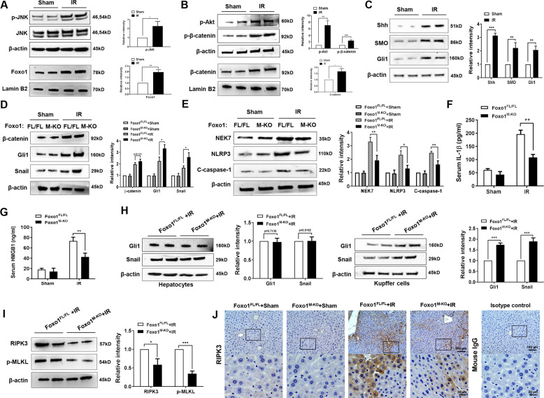
Fig. 2. Disruption of myeloid-specific Foxo1 activates the Hedgehog/Gli1 signaling, and inhibits RIPK3 and NEK7/NLRP3 activation in IR-stressed liver.
Western-assisted analysis and relative density ratio of A p-JNK, Foxo1, B p-Akt, p-β-catenin, β-catenin, C Shh, SMO, and Gli1 in the WT ischemic livers. Western-assisted analysis and relative density ratio of D β-catenin, Gli1, Snail, E NEK7, NLRP3, and cleaved caspase-1 in the Foxo1FL/FL and Foxo1M-KO ischemic livers. ELISA analysis of serum IL-1β (F) and HMGB1 (G) levels in the Foxo1FL/FL and Foxo1M-KO mice after liver IRI (n = 3–4 samples/group). H The expression of Gli1 and Snail was detected in hepatocytes and Kupffer cells by western blot assay. I Western-assisted analysis and relative density ratio of RIPK3 and p-MLKL in the Foxo1FL/FL and Foxo1M-KO ischemic livers. J Immunohistochemistry staining of RIPK3 expression in ischemic livers (n = 4–6 mice/group). Scale bars, 100 and 20 μm. All western blots represent three experiments and the data represent the mean ± SD. *p < 0.05, **p < 0.01, ***p < 0.001.

