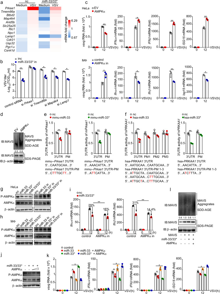
Fig. 5.
miR-33/33* impair the activation of MAVS by targeting AMPK for posttranscriptional silence. a mRNA expression of indicated genes in peritoneal macrophages transfected with nc or miR-33/33* followed by VSV infection (MOI = 1) relative to that of uninfected macrophages transfected with nc. b TCID50 determination of VSV loads in macrophages transfected with negative control inhibitor (inc), miR-33 and miR-33* inhibitors (miR-33/33* in) along with siRNAs as indicated, followed by VSV infection for 12 h. c qRT-PCR analysis of VSV transcripts and Ifnα, Ifnβ, and Isg15 mRNA expression in HeLa cells transfected with empty vector (EV) or expression plasmid for AMPKα (encoded by PRKAA1) (top) or in macrophages transfected with control siRNA (control) or Prkaa1 siRNA (AMPK in) (bottom), followed by infection for the indicated time with VSV (MOI = 1). d SDD-AGE analysis and SDS-PAGE of the aggregation of MAVS in HeLa cells transfected with empty vector (EV) or expression plasmid for AMPKα followed by VSV infection for 12 h. e 3′-UTR luciferase reporter activity of mmu-Prkaa1 in HEK293T cells transfected with nc, mmu-miR-33 or mmu-miR-33*. The normal and mutated mmu-Prkaa1 3′-UTR sequences used are shown below. f 3′-UTR luciferase reporter activity of hsa-PRKAA1 in HEK293T cells transfected with nc, hsa-miR-33, or hsa-miR-33*. The normal and mutated hsa-PRKAA1 3′- sequences used are shown below. g and h Immunoblot analysis of phosphorylated (p-) or total AMPKα in HeLa cells g and macrophages h transfected with nc, miR-33, miR-33*, miR-33 along with miR-33* (miR-33/33*) or inc, miR-33 in, miR-33* in, miR-33 in along with miR-33* in (miR-33/33* in). i qRT-PCR analysis of Ifnα4 and Ifnβ mRNA expression in control and Prkaa1 siRNA (AMPK in)-treated macrophages, which were subsequently transfected with nc or miR-33/33*, followed by infection for the indicated hours with VSV (MOI = 1). j Immunoblot analysis of phosphorylated (p-) or total AMPKα in HeLa cells transfected with nc, miR-33/33*, EV and AMPKα (500–1000 ng/ml) as indicated. k qRT-PCR analysis of VSV transcripts and Ifnα, Ifnβ, Isg15 mRNA expression levels in HeLa cells transfected with empty vector (EV) or expression plasmid for AMPKα along with nc, miR-33, or miR-33* as indicated, followed by infection with VSV (MOI = 1) for the indicated hours relative to those of cells treated with nc plus EV (herein referred to as control). l SDD-AGE analysis and SDS-PAGE analysis of the aggregation of MAVS in HEK293T cells transfected with or without expression plasmid for MAVS, AMPKα along with nc or miR-33/33* as indicated in the figure. *p < 0.05; **p < 0.01 (Student’s t test (b, c, e), one-way ANOVA k or two-way ANOVA i). Data are presented as the mean ± s.e.m. and are representative of three independent experiments.

