Fig. 5.
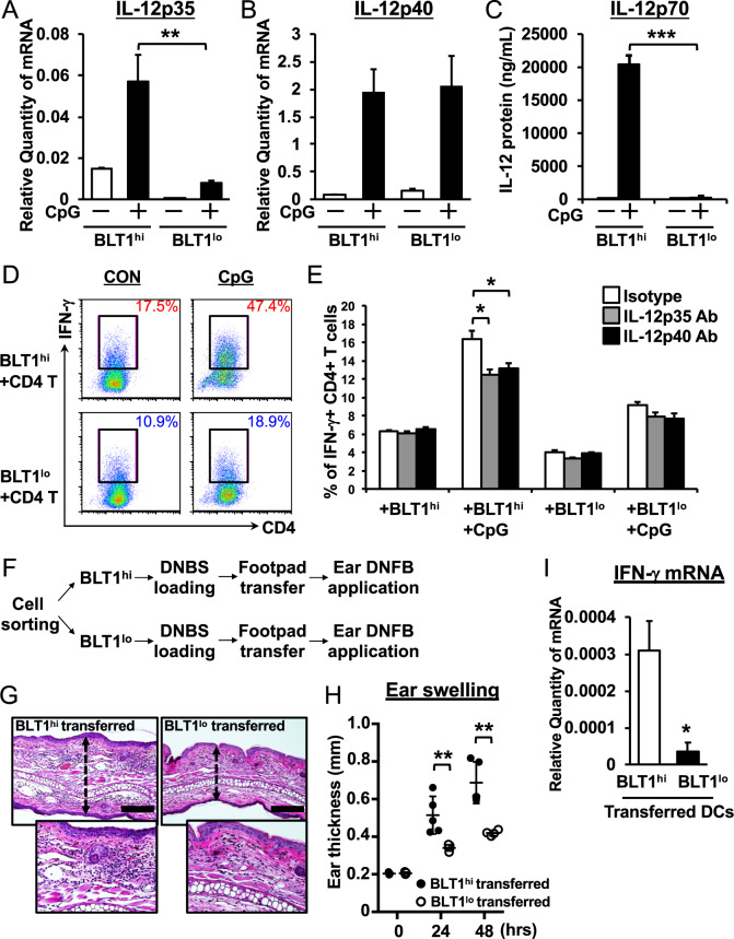
BLT1hi DCs preferentially induce Th1 differentiation by producing IL-12p35. BLT1hi DCs preferentially induce Th1 differentiation by producing IL-12p35. QPCR analysis of IL-12p35 (a) and IL-12p40 (b) expression was performed. Both DC subsets were treated with CpG DNA (500 nM) for 4 h. GAPDH was used as an internal control. c Both DC subsets were treated with CpG DNA for 24 h, and the supernatants were collected. The IL-12p70 protein in the supernatant was quantified by CBA analysis (n = 3; error bars indicate the S.E.M.). d, e Sorted BLT1hi (upper panels) and BLT1lo BMDCs (bottom panels) were cocultured for 3 days with splenic CD4+ T cells derived from OT-II Tg mice (ratio 1:4) in the presence (CpG) or absence (CON) of CpG DNA (500 nM). The cells were stained for intracellular IFN-γ and analyzed by flow cytometry. Cells were incubated for 3 days with neutralizing antibodies specific for IL-12p35 (1 μg/ml) or IL-12p40 (1 μg/ml) to examine the effects of IL-12 on Th1 differentiation. f The schematic shows the experimental flow for DC transfer into the footpad. g, h, i Both DC subsets were loaded with DNBS in vitro for 6 h and then injected into the footpad. Five days later, mouse ear tissue was treated with DNFB, and ear thickness was measured (g). Forty-eight hours after elicitation, ear tissue was collected and stained with H&E (h). Bars, 100 μm. i The mRNA expression of IFN-γ in induced mouse ear tissue was measured by QPCR. β-actin was used as an internal control (n = 3; error bars indicate the S.E.M.). *P < 0.05; **P < 0.01; ***P < 0.001; unpaired Student’s t-test (i); one-way ANOVA with Bonferroni’s post hoc tests (a, b, c, e); two-way ANOVA with Bonferroni’s post hoc test (h)

