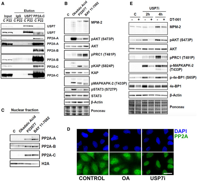
Figure 3. Targeting USP7 inactivates PP2A.

- Immunoprecipitation against IgG, USP7, and PP2A‐C in whole cell extracts of RPE cells that were treated with DMSO (control) or 25 μM P22077 (USP7i). Levels of USP7, PP2A‐A, PP2A‐B, and PP2A‐C are shown. Images from two different exposure times are shown for the better inspection of saturated bands.
- Western blot showing the levels of MPM‐2, pAKT, AKT, pPRC1, pKAP, KAP, pMAPKAPK‐2, pSTAT3, STAT3, and β‐Actin in whole cell extracts of RPE cells treated with DMSO (control), 0.5 μM okadaic acid for 2 h, 25 μM P22077 for 4 h, or 25 μM BAY 11‐7082 for 2 h. Ponceau staining is shown as loading control.
- Western blot showing the levels of PP2A‐A, PP2A‐B, PP2A‐C, and H2A in the nuclear fraction of RPE cells treated with DMSO (control), 0.5 μM okadaic acid for 2 h, 25 μM P22077 for 4 h, and 25 μM BAY 11‐7082 for 2 h.
- Immunofluorescence of PP2A‐A (green) in U2OS cells treated with DMSO (control), 50 μM P22077 (USP7i) for 4 h, or 0.5 μM okadaic acid (OA) for 1 h 30 min. Nuclei were stained with DAPI (blue). Scale bar, 10 μm.
- Western blot showing the levels of MPM‐2, pAKT, AKT, pPRC1, pMAPKAPK‐2, p4e‐BP1, 4e‐BP1, and β‐Actin, in whole cell extracts of RPE cells treated with DMSO (control), 25 μM P22077 (USP7i), and 10 μM of the PP2A activator DT‐061 for 2–4 h. Ponceau staining is shown as loading control.
