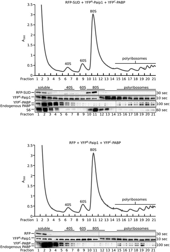
Figure EV2. Polyribosome gradient analysis of lysates from HEK293T cells transiently transfected with either pDEST‐RFP‐SUD/pDEST‐c‐myc‐YFPN‐Paip1/pDEST‐HA‐YFPC‐PABP or pDEST‐RFP/pDEST‐c‐myc‐YFPN‐Paip1/pDEST‐HA‐YFPC‐PABP constructs.

The absorption at 260 nm (A260) and the collected fractions are shown and 40S, 60S, 80S, and polyribosome fractions are labeled. Western blot analysis of the gradient fractions using anti‐RFP, anti‐c‐myc (c‐myc‐Paip1 fusion), anti‐PABP, and rps6 (S6, 40S ribosomal subunit protein) antibodies is shown below.
