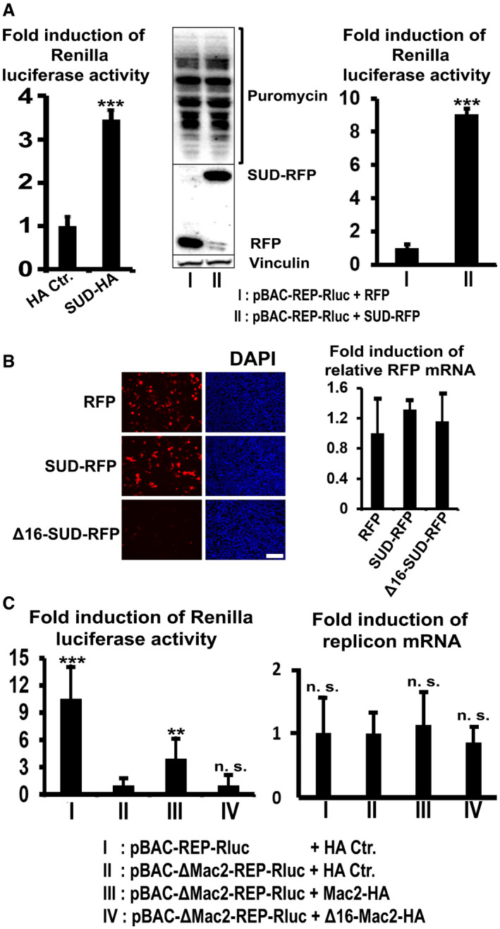
Figure 5. SUD generally stimulates translation but only enhances viral protein synthesis in the replicon‐transfected cells.

- Left image: SUD generally stimulates protein translation level detected by the luciferase‐pcDNA3 reporter (n = 6). Middle image: SUD does not increase the amounts of total protein synthesis (host and viral proteins) in the replicon‐transfected cells. Right image: SUD augments viral protein synthesis (n = 8). HEK‐293 cells growing in 12‐well plates were transfected with the indicated plasmids and replicon DNA. Twenty‐four hours post‐transfection, cells were harvested for Renilla luciferase activity measurement (left and right image). For the ribopuromycylation assay, 24 h post‐transfection cells were pulsed with 3 µM puromycin for 1 h at 37°C before harvesting for Western blot analysis (middle image).
- Δ16‐SUD protein is not detectable but the mRNA level is normal. HEK‐293 cells were transfected with the indicated plasmids. Twenty‐four hours post‐transfection, cells were fixed for DAPI staining (left image) or lysed for a SYBR Green qPCR assay (right image, n = 6). Scale bars represent 100 µm.
- Left image: Mac2 stimulates replication of pBAC‐ΔMac2‐REP‐Rluc replicon, but Δ16‐Mac2 does not (n = 6). Right image: Cellular RNA was isolated for qPCR assay using primers and probes specifically recognizing SARS Nsp14 and β‐actin. The relative pBAC‐REP‐Rluc‐related replicons RNA level was calculated as the ratio of Nsp14 RNA to β‐actin RNA. n = 5.
Data information: ***P value <0.001; **P value <0.01; n. s.: not significant. Data were analyzed using t‐test. Error bars represent SD.
