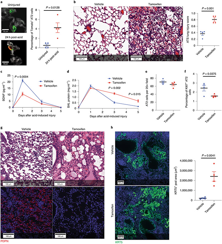
Figure 5 – Loss of AT2 specific BDNF worsens outcomes following sterile and infectious lung injuries.
(A) We quantified tomato positive AT2 cells in BdnfCre-R26TdTomao mice before and 24h after acid-induced lung injury and found a significant increase in co-positive cells following acute lung injury (n=4 mice per group). (B-F) H&E staining and ATS lung injury scores (B), BAL BDNF concentrations (C) BAL protein concentration (D) AT2 cell numbers (E) and number of proliferating AT2 cells (F) of tamoxifen-exposed and vehicle-exposed SftpcCreERT2:BdnfLoxP/LoxP mice 5 days after acid-included lung injury (n= 3 mice per time point for C & D, n=4 mice per group per time point for B, E, & F). (G) H&E and PDPN staining of lung tissue from tamoxifen-exposed and vehicle-exposed SftpcCreERT2:BdnfLoxP/LoxP mice that had been infected with intranasal PR8 influenza (5x10⁻⁵ HAU/mouse) (4 mice per group). (H) KRT5 staining of the mice described in (G) with quantification of KRT5+ pods in both groups (n=4 mice per group). For panels (A), (B), (C), (D), (E), (F) and (H), data is shown as the mean +/− SEM. Statistical significance was determined with a two-tailed Student’s t-test (A), (B), (E), (F), and (H), and with a two-way ANOVA (C) and (D). Scale bars, 100μm (B, G, H) and 10μm (A).

