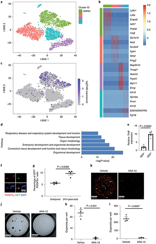
Figure 6 – BDNF-TrkB signaling promotes alveolar epithelial regeneration.
(A-C) Analysis of scRNA-seq of mesenchymal cells from uninjured mice showed that TrkB expression is enriched in mesenchymal alveolar niche cells. (D) Ingenuity pathways analysis show that mesenchymal cells expressing TrkB are enriched with expression of genes that control respiratory system development, organ development and tissue morphology. (E) Expression of TrkB in GFP+ and GFP− cells from TrkBEGFP mice (n=4 mice per group). (F) Expression of TrkB on mesenchymal cells was verified by staining lung tissue form TrkBEGFP mice for PDFRα. (G) Quantification of GFP and PDFRα co-positive cells in TrkBEGFP mice 24 hours after acid-induced lung injury (n=4 mice per group) The gating strategy is shown in Extended Figure 8. (H & I) AT2 cells isolated from a tamoxifen treated SftpcCreERT2-Rosa26mTmG mouse were co-cultured with PDGFRα+ mesenchymal cells for four weeks. ANA-12 abrogated organoid forming efficiency of AT2 cells (n=3 wells per condition). (J & K) The organoid forming capacity of primary human AT2 cells was abrogated by the addition of ANA-12 to the media. Cultures were grown for four weeks. Right-hand panels show average organoid forming efficiency and size from n=3 different donors. For panels (E), (G), (K) and (I), data is shown as the mean +/− SEM. Statistical significance was determined with a two-tailed Student’s t-test (E), (G), (K) and (I). Scale bar, 2.6mm (H, J), 20μm (F).

