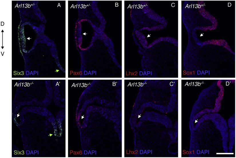
Fig. 3. Optic vesicles are defective in Arl13b−/−.
Immunostaining against Six3 (A, A′), Pax6 (B, B′), Lhx2 (C, C′) and Sox1 (D, D′) in E9.5 embryos. Six3 and Pax6 are classical eye markers whose expression is still detected in the Arl13b−/− OVs and lens placode (A′-B′) although at a lower levels and extension in comparison with Arl13b+/− littermates (A-B). Lhx2 expression is dramatically reduced in the mutant optic vesicles (C′). Sox1, a gene that is not normally expressed in the OVs (D), is abnormally detected in this tissue in the Arl13b−/− OVs (D′). Those results indicate that at E9.5 patterning is affected in Arl13b −/− embryos with the OVs abnormally bending toward the ventral diencephalon. Green arrows indicate the ventral diencephalon (A, A′). White arrows indicate OVs (A- D′). Scale bar: 100 μm. (N: 3).

