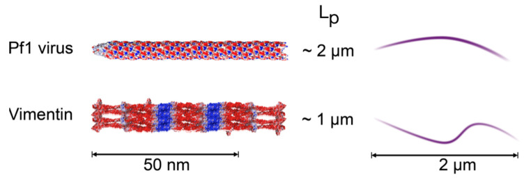
Figure 1.
Schematic diagram of the polyelectrolyte filaments Pf1 virus and vimentin intermediate filaments (VIF). Electrostatic potentials are adapted from ref [1]. Red is negative, and blue is positive. Sketches on the right represent the configuration of 2 µm filaments with the persistence lengths (LP) of these polyelectrolytes.

