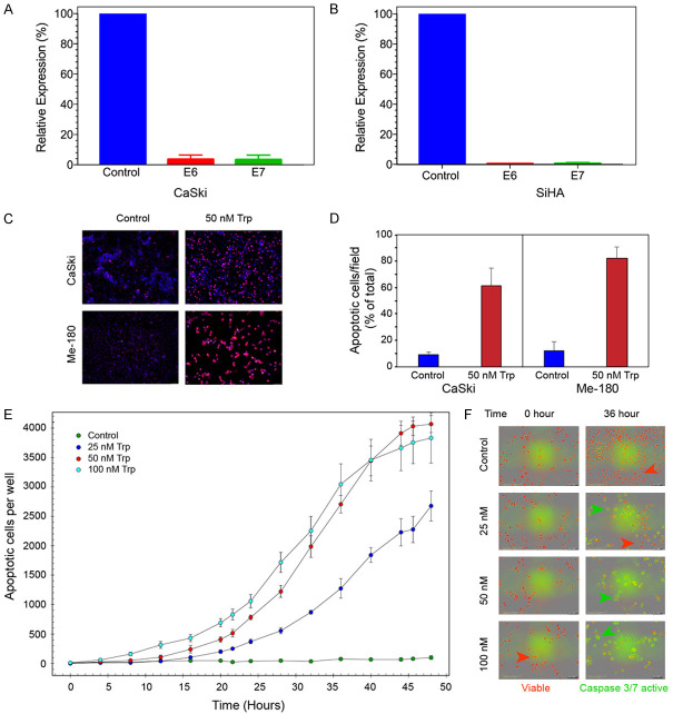
Figure 2.
Triptolide treatment inhibits HPV E6, E7 expression and induces apoptotic cell death. (A) (CaSki) and (B) (SiHa) show the relative levels of E6 and E7 mRNA following treatment with 50 nM Triptolide for 24 hours. mRNA expression was normalized to 18S rRNA. (C) shows representative images of TUNEL assay. Apoptotic cells are labeled red. (D) shows the ratio of apoptotic cells to total number of cells/field. Images were taken 48 hours after treatment with 50 nM of Triptolide. Ten random fields were chosen for image analyses. (E) shows the kinetics (real-time) of concentration dependent Triptolide-induced apoptosis in SiHa cells as determined by InCucyte. Number of apoptotic cells per well was measured using ZOOM software. (F) shows the representative images of control and treated cells before and 36 hours after treatment with Triptolide (Trp). Live cells are labeled with NucRed reagent from InCucyte, and apoptotic cells with active Caspase3/7 are identified by green fluorescence emitted by NucView-488, DNA binding dye which was released from DEVD peptide substrate for Caspase3/7 enzyme.

