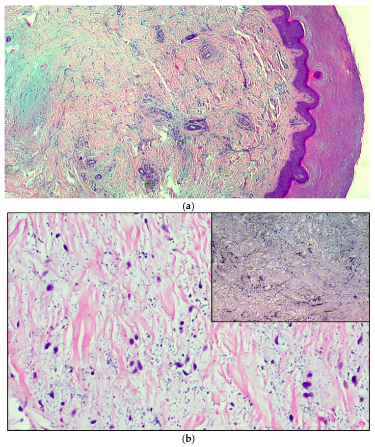
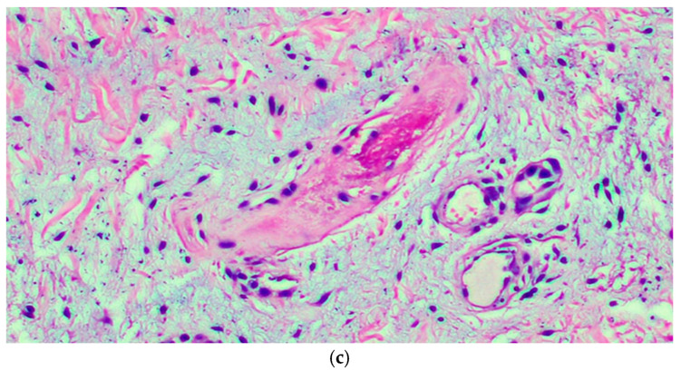
Figure 2.
(a) The microphotograph shows orthokeratosis and mild acanthosis (acral skin), a mild lymphomonocyte infiltrate and myxoid stroma of the dermis. Hematoxylin-Eosin staining, original magnification 100×. (b) In the medial deep dermis, an ample area of edema is evident, with extensive and accentuated fragmentation or even dissolution of the collagen and elastic fibers (the latter confirmed by the Orceina staining), together with the presence of thrombotic foci in the small vessels. (Hematoxylin-Eosin staining, original magnification, 400×, with orcein staining in the insert). (c) Detail of a thrombosed blood vessel together with the presence of fibroblasts of a likely “reactive” significance around extensive, widespread dermal mucinosis. Hematoxylin-Eosin staining, original magnification 400×.


