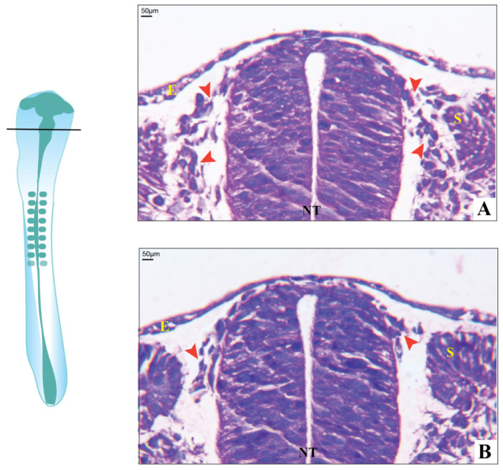
Figure 5.
Histology of chick embryos in the transverse section. (A) Day-2 control with well-developed neural crest cells migrating towards the ventral side of the neural tube (red arrowheads); (B) Day-2 etoricoxib-treated embryo, showing less number of neural crest cells formed along with delayed migration (red arrowheads) (where E—epithelium, NT—neural tube, and S—sclerotome).

