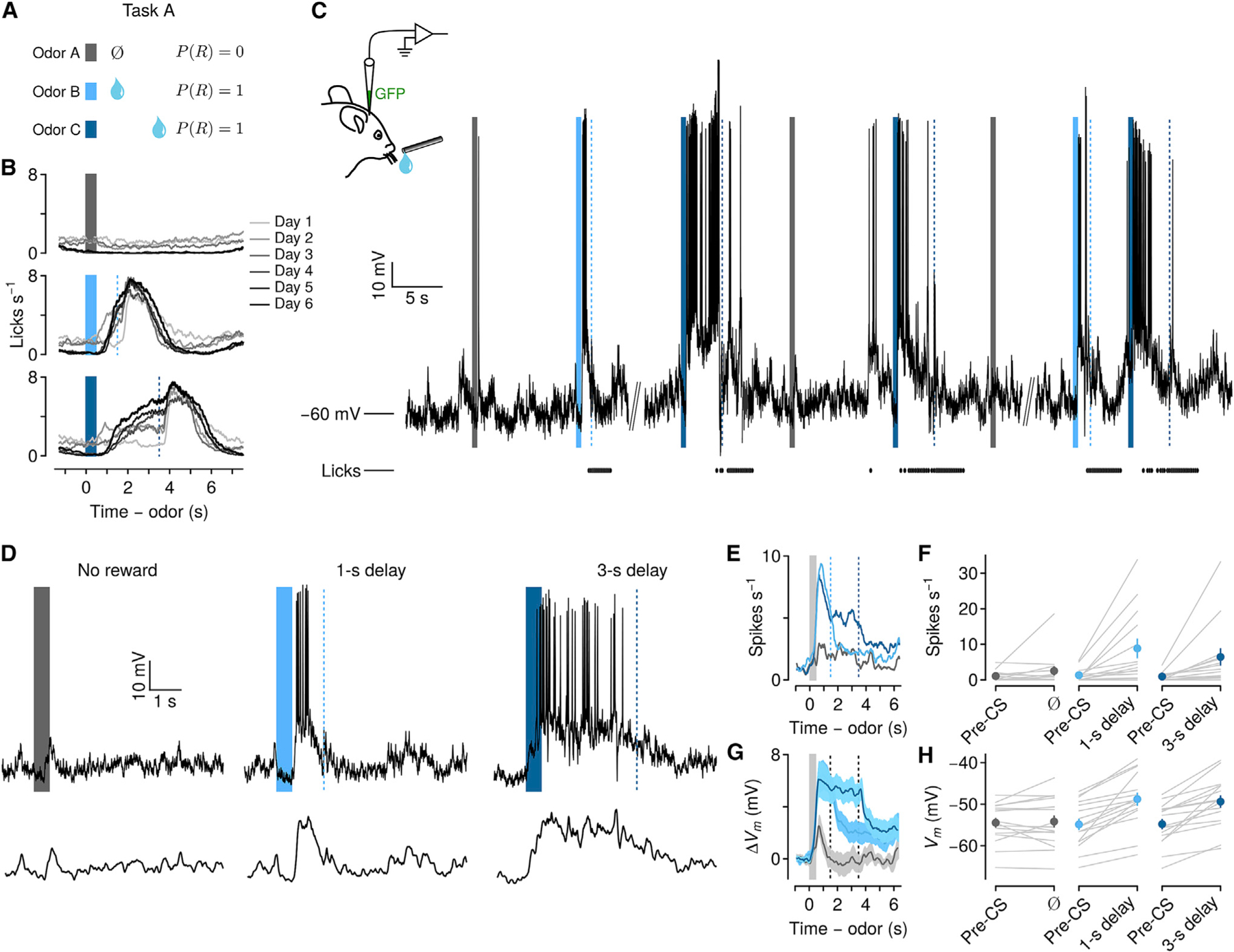
Figure 1. Persistent firing rate and Vm changes in PFC during delays to expected reward.

(A) Behavioral task in which odors predict no reward, reward following a short delay, or reward following a long delay.
(B) Behavioral learning curves show mean lick rates across days of exposure to the task in one mouse. Bars, odor cues; dashed lines, rewards.
(C) Left: schematic of whole-cell recording. GFP plasmid was included in the recording pipette to localize a subset of neurons. Right: Vm from an example neuron over several minutes. Ticks below Vm indicate licks.
(D) Top: example trials of each type from the neuron in (C). Note the persistent increase in spiking and sustained depolarization in the delay between cue and reward. Bottom: Vm with action potentials removed.
(E) Mean firing rates of 15 neurons showing persistent increases during delays to reward.
(F) Firing rates from the same neurons, comparing pre-CS period to delay (individual neurons in gray, points are mean ± SEM).
(G) Mean ± SEM Vm without action potentials from the same neurons.
(H) Vm from the same neurons, comparing pre-CS to delay (individual neurons in gray, points are mean ± SEM).
