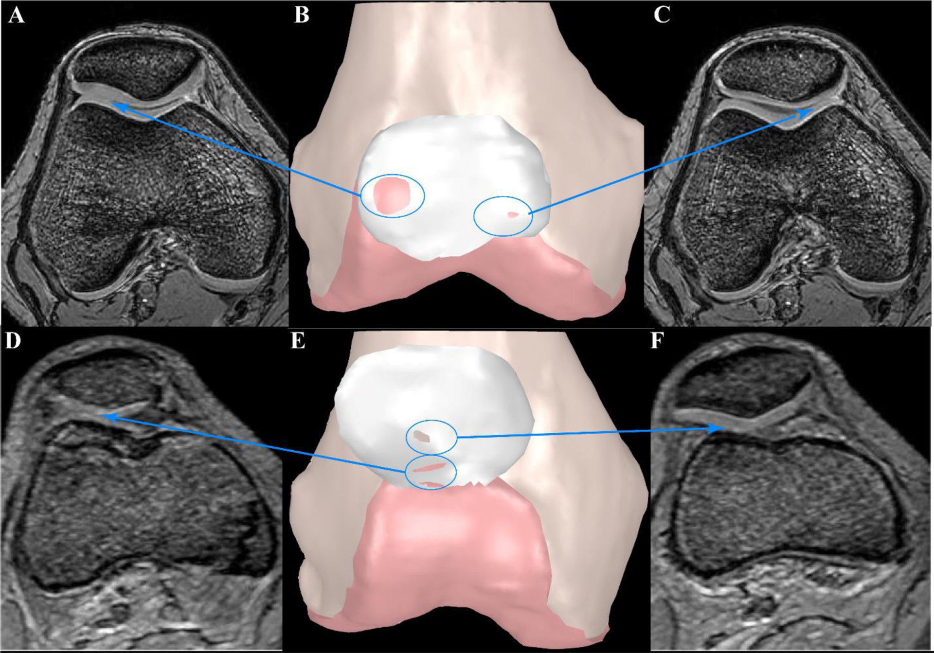
Figure 2. Examples of Patellofemoral (PF) Contact for Two Adolescents with PF Pain.

Static GRE images were used to create 3D bone models (B, E). Femoral and patellar cartilage surfaces were outlined in MIPAV and imported into Geomagic Wrap to create 3D surface models. Contact between surfaces is shown with the underlying surface color appearing on the patellar cartilage. A, C, D, and F represent the axial MR images at the level where contact between surfaces can be visualized. Patient 1 (A-C) demonstrates minimal lateral maltracking (−0.7mm at full extension) with average values (relative to PF pain cohort) for Wiberg index, lateral shaft length, and trochlear-patellar ratio. PF contact is not evenly distributed across the cartilage surfaces. Instead, the contact is concentrated primarily at the anterior-lateral trochlear, with a gap in contact in the central groove. The resulting stress concentration, particular when the quadriceps become active is a likely source of pain. Patient 2 (D-F) exhibits extreme lateral maltracking (−9.5 mm at full extension), patella alta, a short lateral shaft length, and the smallest lateral patellar width. There is cartilage-cartilage contact (D, E) and cartilage-bone contact (E, F). The patella’s path from centered in the groove at deep flexion to extreme lateral maltracking at full extension would foster excessive cartilage sheer forces. Additionally, the femoral shaft provides a force that resists further lateral shift. This combination of sheer force and bone-to-cartilage contact are likely sources of pain.
