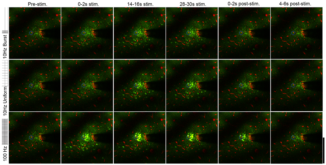
Figure 1.

Two-photon microscopy indicates reveals distinct spatial and temporal responses for neurons with 10 Hz-Burst, 10 Hz-Uniform and 100 Hz stimulation. There is sustained neural activation both close and far from the electrode for 10 Hz-Burst and 10 Hz-Uniform stimulation, while distant cells are only activated during 0–2 s (onset period) for the 100 Hz pattern. Images are presented as averages over the indicated time periods. See supplemental movies 1–3. The Thy-1 GCaMP is colored in green. Vasculature is labeled in red with sulforhodamine 101. The electrode site being stimulated is indicated by the blue dashed circle. Brightness and contrast are unmodified between images. Scale bar is 200 μm.
