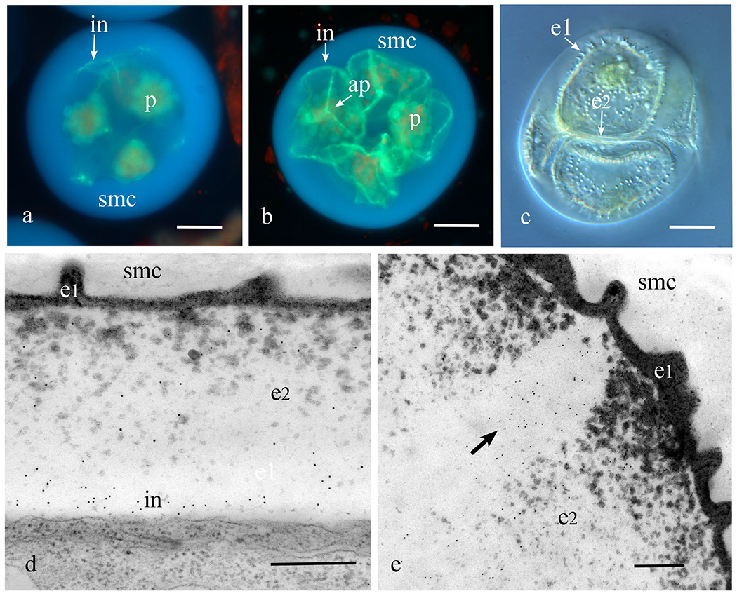
Figure 1. Initiation of the callosic intine.

a-c. Phaeoceros carolinianus. a. Aniline blue fluorescence of a young tetrad enclosed in the spherical spore mother cell (smc) wall showing fluorescence of the callosic intine (in) that develops in patches around the spores; each spore contains a single starch-laden plastid (p). b. Developing spores with single plastids (p) still enclosed in spore mother cell (smc) wall showing newly produced callosic intines (in). The trilete aperture (ap) on the proximal spore surface is enriched in callose. c. DIC image of developing spores enclosed in the mother cell wall showing ornamentation of the outer exine (e1) and inner exine (e2) not yet filled with sporopollenin. Intine is expanding at this stage. d, e. Notothylas orbicularis TEM immunogold labeling with anticallose antibody. e. Developing intine with abundant labels. Outer exine (e1) undulates to form the ornamentation and the inner exine (e2) is filling with sporopollenin. e. Aperture along trilete mark contains callose (arrow points to gold labels) in developing spore wall. Bars: a-c = 20μm, d, e = 2μm.
