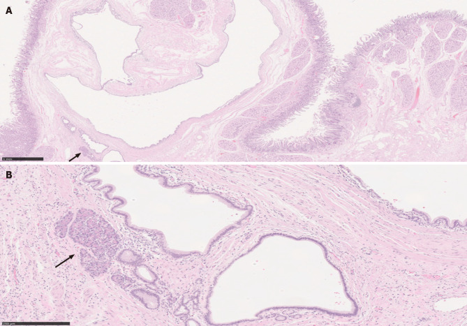
Figure 4.
Paraduodenal pancreatitis. A: This image shows cyst formation, Brunner gland hyperplasia, and ectopic pancreatic tissue (solid arrow) with associated inflammation, consistent with paraduodenal pancreatitis, in a patient with history of significant alcohol use [hematoxylin & eosin (H&E), 20 ×, scale 1 mm]; B: A higher magnification to show the ectopic pancreatic tissue in association with other elements (H&E, 130 ×, scale 250 µm).

