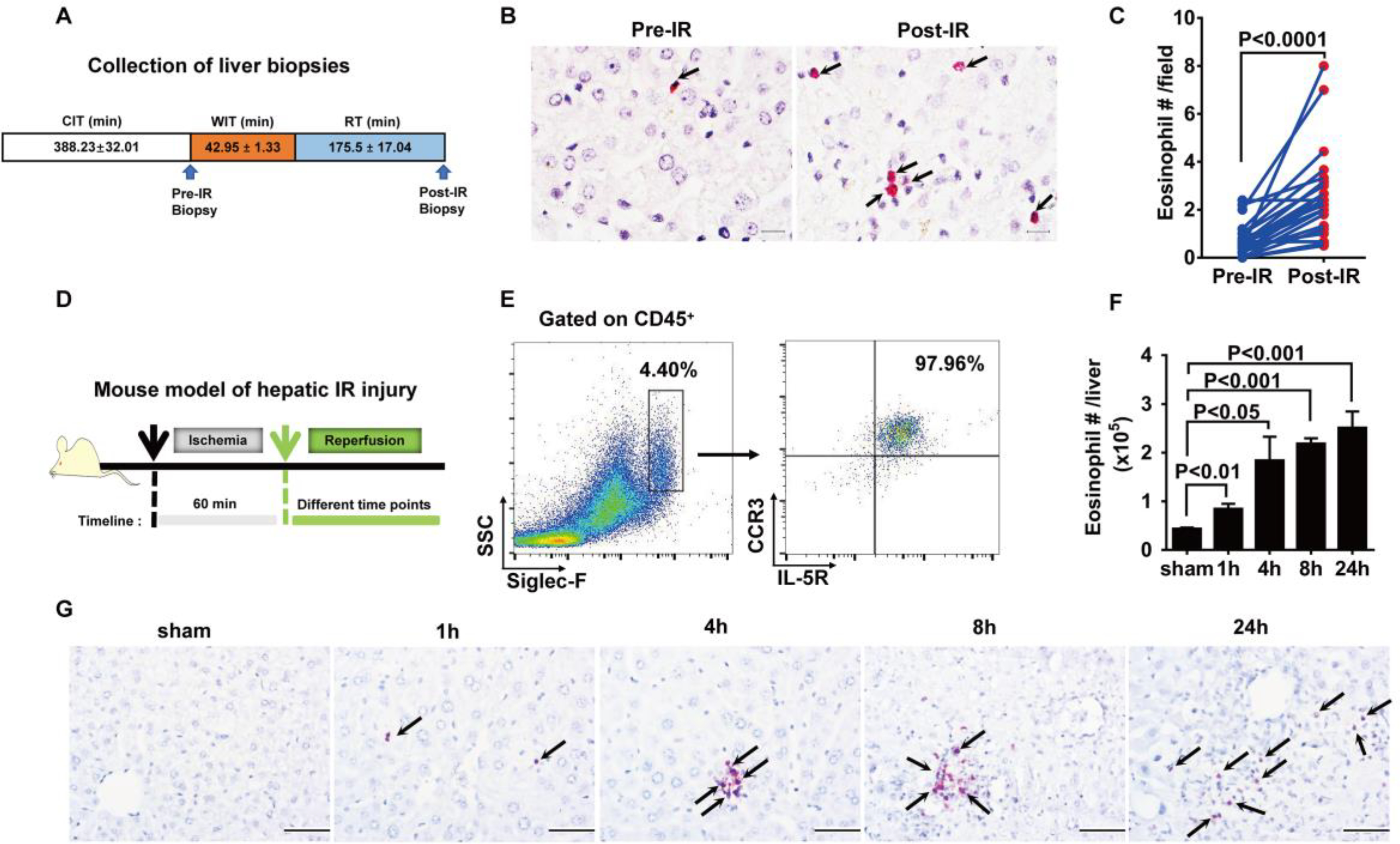
Fig. 1. Eosinophils accumulate in the liver during hepatic IR injury.

(A) Collection of liver biopsies. (B) Eosinophils in human donor liver biopsies (pre-IR) and biopsies from the same tissues after liver transplantation (post-IR) were detected by IHC staining using anti-human EPX antibody. Scale bars, 25 μm. Red indicates EPX staining, and arrows point to EPX-positive eosinophils. (C) The numbers of eosinophils were quantified (n = 22 patients per group). CIT, cold ischemia time; WIT, warm ischemia time; RT, reperfusion time. (D) Male C57BL/6 mice were subjected to hepatic ischemia for 60 min, and the numbers of eosinophils in the liver were measured at 1, 4, 8, or 24 hours after reperfusion. (E) Representative flow cytometry plots and (F) quantification demonstrate the numbers of eosinophils (SSChiSiglec-F+CCR3+IL-5R+cells) in the liver after IR injury (n = 4 per group). SSC, side scatter. (G) IHC staining of eosinophils using anti-mouse MBP antibody. Arrows point to MBP-positive eosinophils stained red. Scale bars, 200 μm. Images shown are representative of n = 4 mice per time point. A two-tailed paired Student’s t test with Welch’s correction was performed in (C). A one-way ANOVA was performed in (F).
