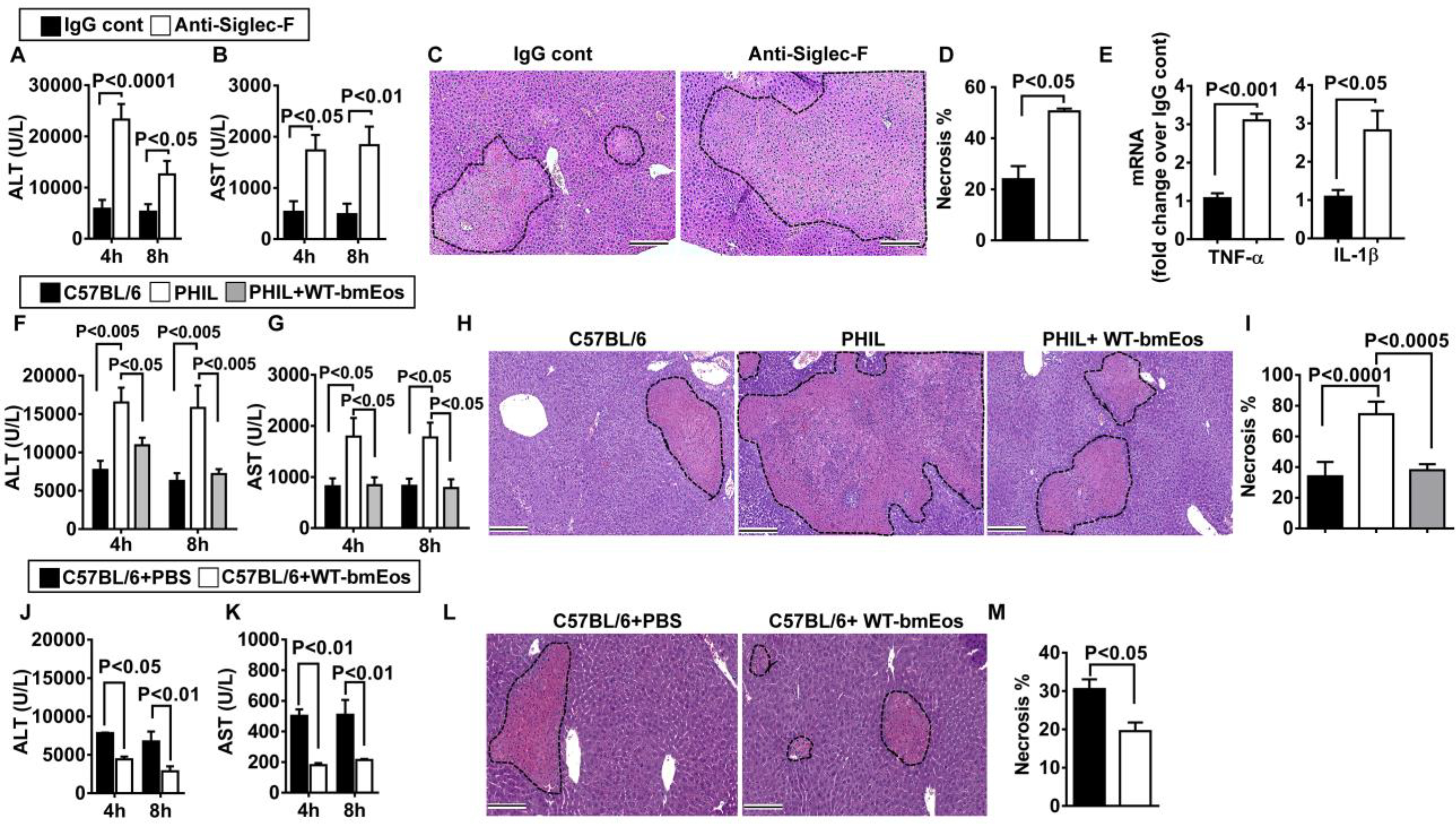
Fig. 2. Eosinophils protect against hepatic IR injury.

(A to E) Eosinophils were depleted by anti–Siglec-F antibody in male C57BL/6 mice, and control mice were treated with IgG (n = 5 in IgG control and n = 3 in anti–Siglec-F treatment group), and serum concentrations of ALT (A) and AST (B) were measured at 4 and 8 hours after reperfusion. (C) Liver necrosis (scale bars, 200 μm) was evaluated and quantified (D) at 24 hours after reperfusion. (E) mRNA expression of cytokines in liver nonparenchymal cells was measured at 4 hours after reperfusion (n = 6 mice per group). (F to I) WT littermates, eosinophil-deficient PHIL mice, and PHIL mice receiving WT-bmEos (2 × 10⁷) were subjected to hepatic IR injury (n = 4 mice per group). Serum concentrations of ALT (F) and AST (G) were measured at 4 and 8 hours after reperfusion. (H) Liver necrosis (scale bars, 200 μm) was examined and quantified (I) at 24 hours after reperfusion. (J to M) WT C57BL/6 mice (n = 4 mice per group) received WT-bmEos or PBS as a control. Serum concentrations of ALT (J) and AST (K) were measured at 4 and 8 hours after reperfusion. (L) Liver necrosis (scale bars, 200 μm) was examined and quantified (M) 24 hours after reperfusion. A two-tailed unpaired Student’s t test with Welch’s correction was performed in (D), (E), and (M). A one-way ANOVA was performed in (I). A two-way ANOVA was performed in (A), (B), (F), (G), (J), and (K).
