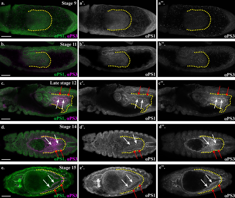
Figure 7.
αPS1 and αPS3 integrin subunits become expressed in the midgut during MET and localize to distinct domains.(a–e) Wild-type embryos stained for αPS1 (green) and αPS3 (magenta). White arrows indicate αPS3 in the apical domain of the midgut cells, and red arrows point out αPS3 in the basal region. Yellow dotted lines outline the posterior midgut. Scale bars, 50 µm.

