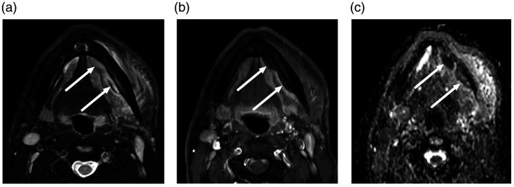
Fig. 4.
Submandibular space subperiosteal abscesses. A 32-year-old man presented with left submandibular space swelling and subsequent dental extraction. Axial MRI slices (a, T2-weighted Dixon water; b, gadolinium-enhanced T1-weighted Dixon water; c, apparent diffusion coefficient from diffusion-weighted imaging) demonstrated non-enhancing collections with restricted diffusion surrounded by abnormal tissue enhancement (arrows) on the medial aspect of the body of the mandible, consistent with subperiosteal abscesses.

