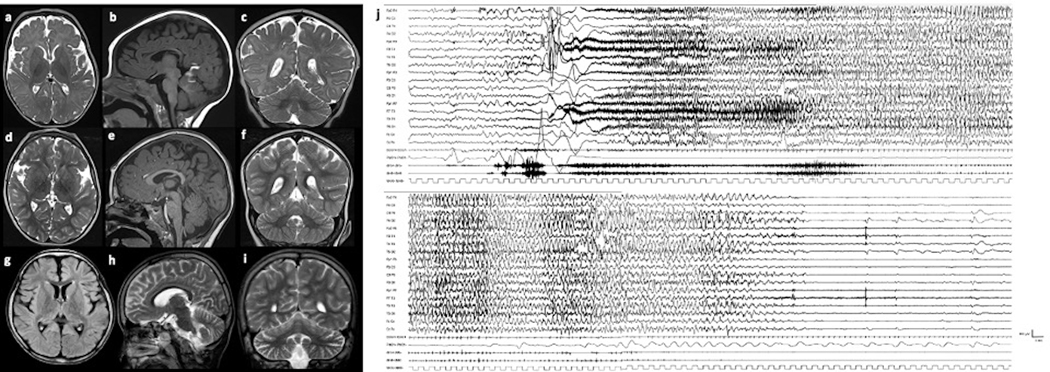
Figure 1. Brain MR of patients #L and #I (a-i), and Ictal EEG of patient #L (j).
Patient #L at the age of 2 years 3 months (a-c) and 2 years 7 months (d-f): cerebral atrophy, with enlarged subarachnoid spaces around the frontal and insular lobes, without significant progression of the atrophy. Patient #I at the age of 8 years (g-i): mild cerebral and cerebellar atrophy with enlarged subarachnoid spaces around the frontal and insular lobes and cerebellar folia. Ictal EEG of patient #L at the age of 42 days (j). Ictal discharge starts with diffuse bilateral, symmetrical, low-voltage fast activity, increasing in amplitude and decreasing in frequency. The patient has a massive tonic contraction with perioral cyanosis and sialorrhea. Polygraphic recording shows ictal bradycardia at seizure onset for about 5 seconds concomitant with the beginning of the tonic phase (see bilateral contraction of upper limb in deltoids). Afterwards, the patient appears floppy and pale associated with a brief compensatory tachycardia. The seizure spontaneously ends after 86 seconds.

