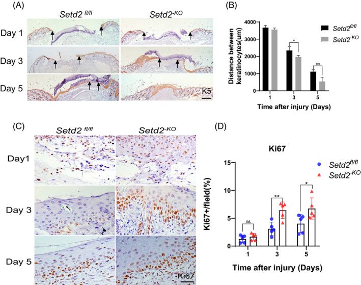
FIGURE 3.

Setd2 deficiency leads to accelerated KC proliferation and migration during wound healing A, K5 staining (brown) of wounds in Setd2fl/fl and Setd2‐KO mice at the specified time points after injury. Arrows indicate the edges of migrated KCs. Scale bars: 500 μm. B, Quantification of the distance between the KCs in the wounds of Setd2fl/fl and Setd2‐KO mice; n = 5 wounds/group. C, Immunohistochemical staining using anti‐Ki67 antibody performed on Setd2fl/fl and Setd2‐KO wound sections collected on days 1, 3 and 5. Representative images. Scale bars: 50 μm. D, Quantification of Ki67‐positive KCs; data are presented as the percentage of total KC number; n = 5 wounds/group. Data are presented as the mean ± SD; and statistical significance was determined using a two‐tailed Student's t test; n.s, no significance, *P < .05, **P < .01 and ***P < .001
