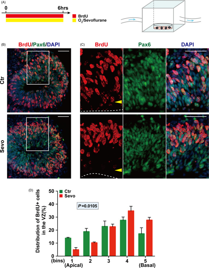
FIGURE 3.

Sevoflurane exposure impairs the INM of RGPs in human cerebral organoids. (A) Schematic diagram of the timing of sevoflurane exposure and anaesthetic apparatus for cerebral organoid at day 30. (B) Representative images of sections from cerebral organoid after treatment stained with Pax6 and BrdU at day 30. (C) The surface of the VZ‐like structure is outlined by a dashed line. (D) Quantification of the distribution of BrdU+ cells in each bin. The VZ‐like structure determined by Pax6 + cells in the cerebral organoid section was divided equally into 5 bins. n = 3 for each group. Data are presented as mean ± SEM. Scale bars represent 50 μm
